 3899 8999


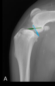
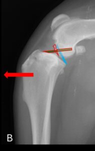
The stifle (knee) joint is stabilized by two primary ligaments: the cranial cruciate ligament (shown in green in image A) and the caudal cruciate ligament (shown in blue in image A). The cranial cruciate ligament’s main role is to prevent the tibia from moving forward relative to the femur, while the caudal cruciate ligament prevents backward displacement of the tibia. Cranial cruciate ligament rupture can occur due to trauma (similar to injuries seen in skiers or football players) but is more commonly the result of a gradual degenerative process. When this ligament ruptures (image B), the tibia moves abnormally forward during weight-bearing, stretching the joint capsule and causing pain.
HOW IS A CRANIAL CRUCIATE LIGAMENT DISEASE /RUPTURE DIAGNOSED
The diagnosis of cranial cruciate ligament rupture is primarily made through an orthopaedic examination, during which the stifle joint is palpated, and specific tests are conducted to assess its stability. The diagnosis can be more challenging if the ligament is only partially ruptured or if the injury occurred long ago. In these cases, examination under sedation and radiographs are often necessary to detect indirect signs of ligament disease, as the cruciate ligament itself is not visible on X-rays. In some instances, a definitive diagnosis is achieved through arthroscopy
WHAT ARE THE TREATMENT OPTIONS FOR CRUCIATE LIGAMENT DISEASE
- Medical management
While medical management can help alleviate your pet’s discomfort and may lead to some improvement, it generally does not result in a good long-term outcome. Regardless of your pet’s size, full recovery is unlikely, and most animals will continue to experience some degree of impairment.
- Surgical Management:
Regardless of the surgery technique used. A small arthrotomy (joint incision) is performed as part of the surgery. It is to assess the menisci (two shock absorber pads between the femur and the tibia) that can be injured following the cranial cruciate ligament rupture. When a lesion or a tear is identified, the lesion is resected. Failure to identify a lesion may lead to some degree of persistent lameness.
- Surgical Management: Lateral Fabellar Suture
This technique was widely used several decades ago and involves placing a prosthetic suture on the lateral side of the stifle joint to act as a temporary ligament. The suture provides stability while surrounding tissues form scar tissue, which ultimately stabilizes the joint. Over time, implant failure is expected, and the joint’s stability relies on the strength of the scar tissue. If the implant fails before sufficient scarring occurs, it may result in prolonged or suboptimal recovery. Dogs often experience a longer recovery period with this technique, and recent studies have shown that osteotomy techniques, such as TPLO, yield superior short and long-term outcomes, particularly in medium to giant-breed dogs, which may experience some long-term lameness with the lateral fabellar suture technique. Nonetheless, the lateral fabellar suture is still performed because it is more cost-effective and requires less surgical expertise and specialized instrumentation compared to TPLO.
- Surgical Management: TPLO (Tibial Plateau Leveling Osteotomy)
- TPLO: What is it?
Osteotomy procedures take a different approach compared to traditional ligament repair techniques (lateral fabellar suture). Rather than attempting to replace the damaged ligament with a prosthesis that will eventually fail, the osteotomy alters the joint’s anatomy to eliminate the need for a functioning cranial cruciate ligament altogether. The procedure involves cutting and rotating the shin bone (tibia) to flatten the slope of its top surface (tibial plateau), thereby preventing abnormal movement of the femur relative to the tibia (images C-D). The cut bone is then stabilized with a metal plate and screws (images E-G) to hold it in the new position while healing occurs. These implants typically remain in place long-term unless complications, such as infection, arise and require removal.
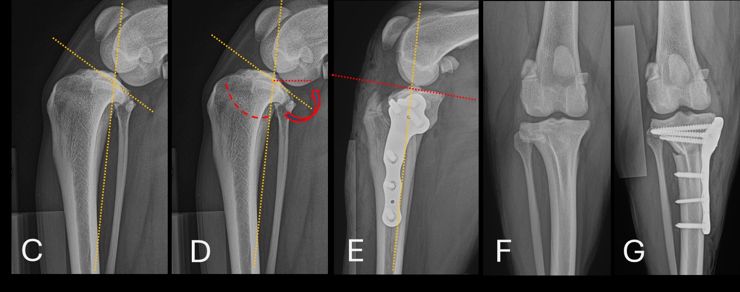
During TPLO, a curved incision is made in the tibia (Image D), allowing for precise rotation and leveling of the tibial plateau. This modification changes the biomechanics of the joint, making the femur less likely to slide down the tibial plateau during weight-bearing. As a result, the stability of the stifle joint is significantly improved. This technique is associated with a quicker recovery and better long-term outcomes. This is the technique recommended by AMAH surgery service
- TPLO: Potential Complications
Postoperative complications can include wound swelling or infection, implant failure, fractures, late meniscal injury (damage to the joint’s shock absorber), patellar tendon inflammation, progression of osteoarthritis, and persistent lameness. The overall complication rate is approximately 5-8%. Strict rest and diligent post-operative care at home are crucial to achieving a successful outcome. Major complications, such as fractures or implant failure, are often the result of inadequate rest during the recovery period
- TPLO: Post-Operative Care
Post-operative care involves strict rest for a period of 6 to 8 weeks. Your pet will be sent home with pain medications and, if necessary, antibiotics. It is essential to prevent your pet from running or jumping during this time. They should be confined to a crate, cage, or small pen, and if that’s not possible, they should at least be kept on a leash and restricted to non-slip surfaces. Short, controlled leash walks of 5 to 10 minutes, 3 to 5 times a day, are permitted for bathroom breaks. If feasible, you may perform gentle physiotherapy exercises (mainly passive range-of-motion activities) at home. Your dog should be consistently using the operated limb one week after surgery, and this should improve further over the coming weeks.
Following the post-operative instructions closely is critical, as failure to do so can lead to complications such as implant failure or bone fractures. After 6 to 8 weeks, your pet will be re-examined, and follow-up radiographs will be taken to assess bone healing. Mild sedation may be required for this visit to ensure precise imaging. If the bone is healing as expected, your pet can gradually begin to resume normal activity
- TPLO: What outcome should I expect?
Approximately 95% of patients report good to excellent outcomes following TPLO surgery. A recent study found that the limb function of the operated leg in dogs who underwent TPLO was comparable to that of healthy dogs during both walking and trotting within 6 to 12 months post-surgery, demonstrating superior results compared to other techniques included in the study. Although it is rarely clinically significant, osteoarthritis can continue to develop in the joint after surgery. Therefore, it is recommended that all dogs receive management strategies for osteoarthritis treatment. This includes dietary management, as obesity increases the risk of contralateral cruciate disease, progression of osteoarthritis, and potential surgical complications
- Is TPLO Suitable for Animals of All Sizes?
Yes, TPLO surgery has been associated with good to excellent outcomes in dogs and cats of all sizes. We have the appropriate implants to perform the procedure on both giant breed dogs and miniature patients.